3.6 Animal eyes⧉
Human vision is one solution among many, and a parochial one. Eyes have evolved independently dozens of times, and surveying the alternatives is the best cure for treating our own visual system as the definition of "seeing." A few questions organize the tour, the same ones that organize the whole book's treatment of a camera: how the image is formed (optics), how the receptor array is laid out and how faint or fine a signal it can resolve (sensitivity and acuity), and how color is sensed (the opsins and their wiring). The optical side connects back to Image formation; the color side extends the opsin story of Perceptual color and trichromatic vision.
3.6.1 The optics: many ways to form an image⧉
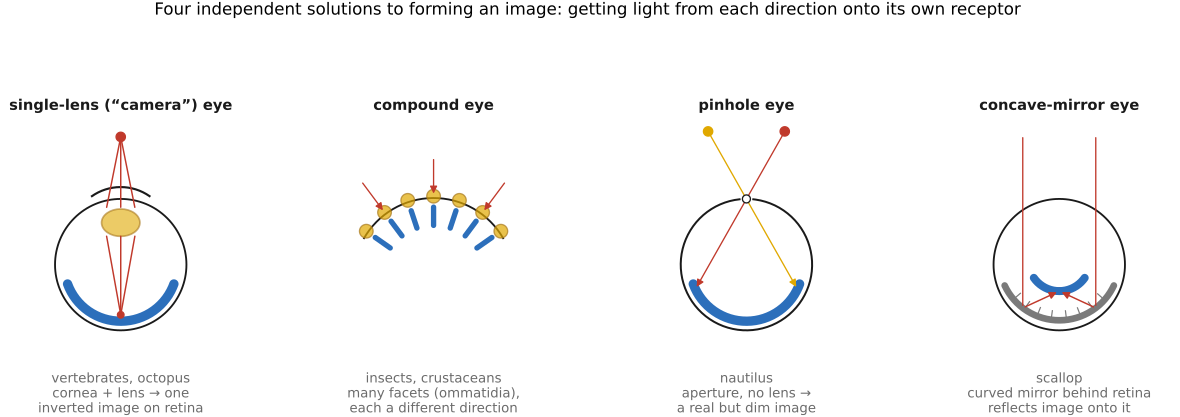
Forming an image means getting light from each direction in the scene onto a different receptor, the same plenoptic-sampling problem a camera solves with a lens. Nature has found several radically different answers (Figure 3.6.2).
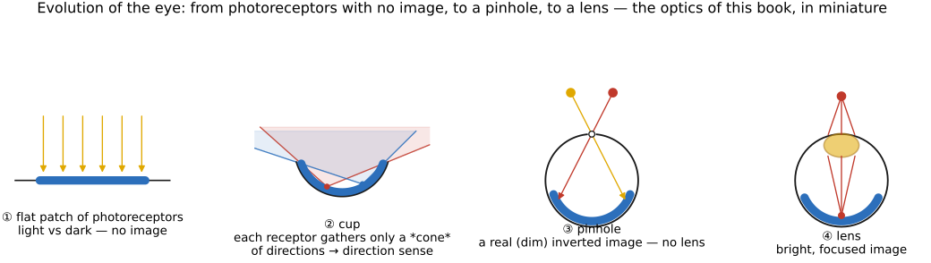
Before the variety, the sequence. A full eye is not an all-or-nothing leap but a ladder of small, individually useful improvements (Land & Nilsson Land & Nilsson, Animal Eyes). Start with a flat patch of photoreceptors that registers only light versus dark; fold it into a cup, and the rim shades part of the sheet, giving a crude sense of direction; close the cup to a pinhole, and a real (if dim) inverted image forms with no lens at all; finally fill the aperture with a lens for a bright, focused image. That progression (from no image to a focused one) recapitulates the optics of this book in miniature (Figure 3.6.1).
Even the bottom rung has a subtler cousin. The single-celled Euglena has no eye at all, only a light-sensitive spot shaded by an adjacent speck of orange pigment, its stigma. As the cell corkscrews through the water the pigment speck sweeps past the receptor and periodically shadows it, so the receptor reads the direction of the light from the rhythm of its own shadow, and the organism steers toward it. The trick is the exact optical inverse of a pinhole: where a pinhole is a small hole that lets a pencil of light through, this is a pinspeck, a small opaque speck that blocks one and casts a shadow, and a shadow carries directional information just as an image does. It is the same anti-pinhole principle we later exploit to form accidental images from ordinary occluders (the accidental pinspeck).
Read as engineering, each design is a point on a handful of tradeoffs, and evolution is the optimizer that found it under an animal's particular constraints: the space the eye is allowed (a fly's head cannot hold a human eyeball), the light of its environment (a bright reef, the deep sea, a night forest), the task it must serve (spot a predator on the horizon, track a darting fly, judge the distance of a pounce), and the processing budget behind the retina, since more receptors mean more data for a nervous system to carry and interpret. No design wins outright; each spends those currencies differently.
- Single-lens ("camera") eyes, like ours and the octopus's, use one refracting element to cast a single inverted image on a sheet of receptors, the design we have been modeling all chapter. It delivers the highest resolution per eye, but it is costly on every axis: a single large aperture, a bulky spherical eyeball, and a retina of millions of receptors that only a large, expensive brain can read, which is why the sharpest camera eyes belong to animals that can pay the neural bill. The vertebrate and cephalopod versions evolved separately and even wire their retinas back-to-front relative to each other, a textbook case of convergent evolution onto the same optics.
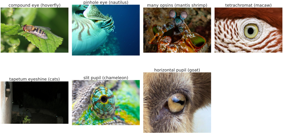
- Compound eyes (insects, crustaceans) tile the visual field with hundreds or thousands of ommatidia, each a tiny lens feeding one or a few receptors and pointing in a slightly different direction. Resolution is set by how finely the facets are packed, not by a single aperture, so acuity is modest, but the field of view is enormous and the temporal response very fast (one reason flies are so hard to swat). It is, in effect, a fixed array of one-pixel cameras. Its tradeoff is driven by space: a compound eye stays tiny and light, wraps almost all the way around a small head, and answers fast on little neural hardware, but facet size and diffraction cap its resolution so hard that matching human acuity would take an eye roughly a meter across. That single fact is why a small, fast, wide-viewed insect took this branch and a large, sharp-eyed vertebrate did not.
- Lens-less designs solve image formation without refraction at all: the pinhole eye of the nautilus (an aperture with no lens, sharp but dim, exactly the pinhole-camera tradeoff from Image formation), and the mirror ("reflecting") eyes of scallops and some deep-sea crustaceans, which focus by a curved multilayer mirror behind the retina rather than a lens, the biological analogue of a catadioptric camera. Both are cheap, compact answers for animals whose goal is sensing change or coarse shape rather than fine detail: the pinhole needs no lens to grow, feed, or focus, and the mirror folds real focusing power into a thin, flat eye that suits a scallop's narrow shell gape or the deep dark, where a refracting lens would lose too much light.
- Eyes built for a niche. Many designs are tuned to a task more than to general acuity: the tapetum lucidum, a reflective layer behind the retina in cats, dogs, and deer, bounces unabsorbed photons back through the receptors for a second chance, boosting night sensitivity at the cost of a little blur (and causing eyeshine in a flash photo). Deep-sea fish stack receptors or grow tubular eyes to wring signal from a few photons; the four-eyed fish Anableps splits its cornea to focus above and below the waterline at once. Each of these spends resolution, or bulk, or optical simplicity to win the one thing its world demands, which is the recurring shape of every eye in this chapter: not a single best design, but a budget spent to fit a niche.
3.6.2 The pupil: aperture shape as a clue to ecology⧉
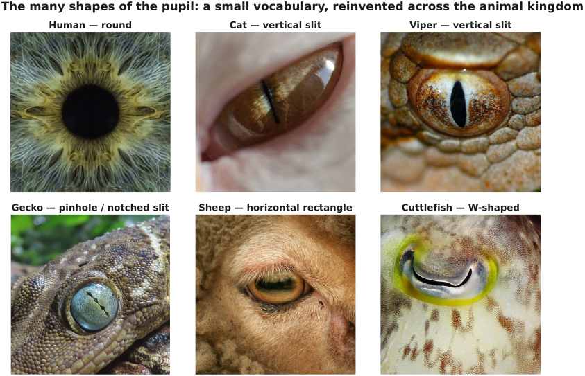
The pupil is the eye's aperture, the biological f-stop that meters how much light reaches the retina and sets the depth of field. Across animals its shape is far from incidental: it tracks how a creature makes its living so tightly that you can often guess an animal's niche from the pupil alone (Banks and colleagues, 2015) (Figure 3.6.4).
Round pupils, ours and the lion's and most birds', are the simplest case: a symmetric aperture that constricts concentrically and blurs out-of-focus points by the same amount in every orientation. They dominate among animals that forage actively in daylight, and among tall predators whose eyes sit well above the ground.
Vertical slit pupils, in cats, foxes, geckos, and many snakes, mark ambush predators that hunt across both bright day and dark night. The slit buys two things. First, an enormous operating range: it can clamp down to a barely-open sliver and dilate to a wide round hole, changing its area by a factor of a hundred or more where a round pupil manages a fraction of that, so one eye can work from noon glare to near-darkness. Second, a depth cue tuned for the ambush. A vertical slit blurs vertical edges with distance while keeping horizontal edges and horizontal binocular disparity sharp, so an animal lying low to the ground and striking at close range reads distance from the interplay of defocus blur and stereopsis. The vertical orientation lines up with the upright contours (grass, stems, trunks) that carry the strongest distance information.
Horizontal, rectangular pupils belong to grazing prey such as goats, sheep, horses, and deer. A wide horizontal slit stretches the field of view into a panoramic strip along the ground and the horizon, exactly where a predator will appear, while cutting the glare from the bright sky overhead and giving a sharp, well-exposed image of the terrain underfoot. Their most striking trick is that the eyes counter-rotate as the head lowers to graze (cyclovergence), keeping the slit aligned with the horizon so the panorama stays level whatever the head is doing.
W-shaped and U-shaped pupils appear in cuttlefish, whose constricted pupil in bright light is a wavy W. One idea is that it balances the uneven light of a scene that is bright above and dim below; another, more speculative, is that because cuttlefish are colorblind the wide, oddly-shaped aperture lets them read color indirectly from chromatic aberration, inferring wavelength from how sharply a scene comes to focus (Stubbs and Stubbs, 2016).
Pinhole and multiple-aperture pupils are the nocturnal extreme. Some geckos close their slit not to a line but to a row of separate pinholes; each pinhole forms its own slightly shifted image, and the offset between the overlapping images is a strong cue to distance, while the tiny apertures keep the image sharp in daylight for an eye built for the dark.
The moral rhymes with the rest of the chapter. The aperture we reduce to a single number when we set an f-stop is, across the animal kingdom, a shape, and that shape is tuned to a way of life: circles for active day-foragers, vertical slits for ground-hugging ambush hunters, horizontal bars for wary grazers.
3.6.3 Amphibious vision: focusing in air and in water⧉
An eye optimized for air is hopelessly out of focus underwater, and the culprit is the cornea. In air most of an eye's focusing power comes not from the lens but from the front surface of the cornea, where light crosses the large index step from air (about 1.0) into tissue (about 1.38). Submerge that cornea and it is now bounded by water (about 1.33), almost its own index, so the corneal step nearly vanishes and most of the eye's power goes with it. A person who opens their eyes underwater turns strongly farsighted for exactly this reason, and goggles cure it only by restoring a flat pocket of air in front of the cornea. Any animal that must see in both media faces the same bind: the workhorse refracting surface works in air alone.
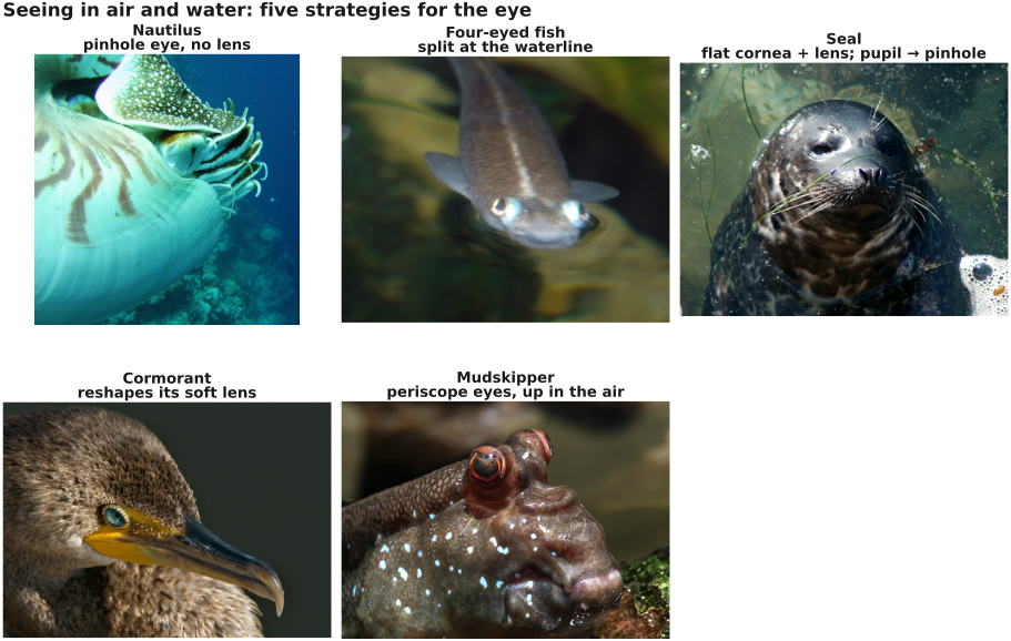
Evolution has answered this several ways (Figure 3.6.6), and you can trace each one through a schematic eye (Figure 3.6.5).
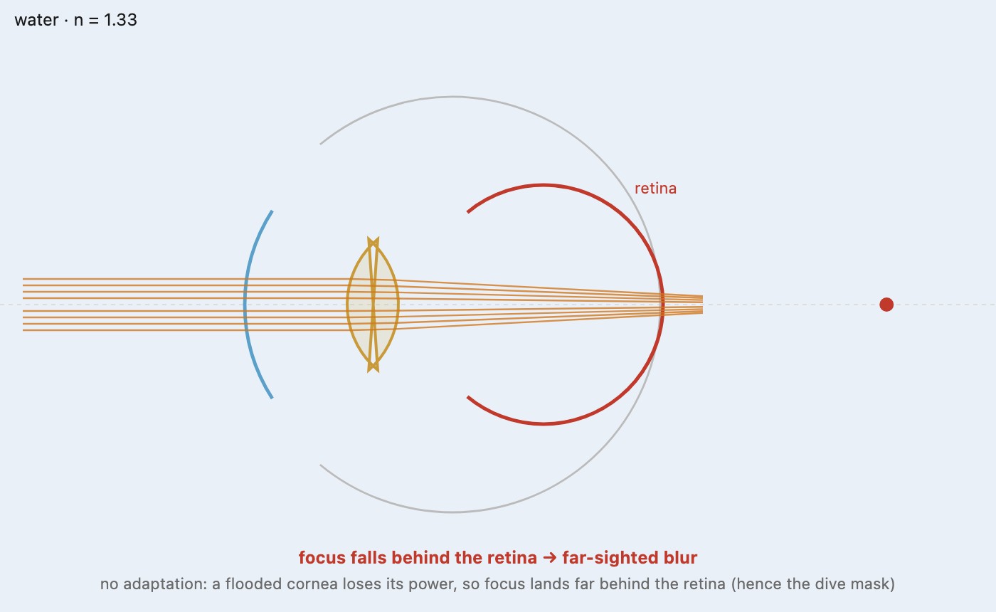
Sidestep refraction with a pinhole. The simplest answer refuses to refract at all. A pinhole forms its dim, inverted image by pure geometry, rays selected through a small aperture, so it owes nothing to the index of the surrounding medium and works identically in air and in water. The nautilus's lens-less pinhole eye (§2.4.1) is fully aquatic, but the same medium-independence is why stopping a pupil down toward a pinhole helps an amphibious eye cope in its wrong medium: a tighter aperture cannot restore the corneal power a flooding took away, yet it deepens the depth of field enough to keep the out-of-focus world tolerably sharp, at the price of the light a pinhole always spends. It is less a full solution than an escape hatch, which is why the animals below mostly fix the refraction instead.
Do all the focusing with the lens. Fully aquatic eyes give up on the cornea and focus with a near-spherical, high-index lens, the trick a fish uses. Seals and sea lions carry it into an amphibious life with a nearly flat cornea, which has little power in either medium, paired with a strong spherical lens that focuses underwater where they hunt. In air they would be badly myopic, so the pupil closes to a tiny pinhole in bright light, trading light for the depth of field that keeps the aerial world acceptably sharp.
Split the eye in two. The four-eyed fish Anableps swims at the surface with the waterline drawn across the middle of each eye. Its cornea is divided into an upper half that refracts air and a lower half bathed in water, and its lens is egg-shaped, thicker along one axis than the other, so light entering from above and from below travels different optical paths and both land in focus on the retina. One eyeball runs two optical systems at once, watching for predators above the surface and prey below it.
Reshape the lens on demand. Amphibians, turtles, and above all diving birds focus by deforming the lens rather than the cornea. Cormorants, dippers, and mergansers have exceptionally soft lenses and powerful ciliary muscles that squeeze the lens through the ring of the iris as they dive, adding tens of diopters of power to replace what the flooded cornea lost. Some diving birds add a clear refracting window in the nictitating membrane, a third eyelid that doubles as a contact lens underwater.
Or cross the waterline the other way, to see in air. The mudskipper does the reverse of the others: a fish that spends hours out of the water on tropical mudflats, it lifts bulging, periscope-like eyes into the air and sees there with a flattened cornea that, back in its refracting medium, once again has an index step to work with. The amphibious problem is symmetric, and evolution has met it from both banks.
The through-line is the one that runs through all of the eye's optics: there is no single design best in every condition, only solutions matched to an animal's world. An amphibious eye simply has two worlds to satisfy at once, and each answer, the flat-cornea seal, the split-eyed Anableps, the lens-pumping cormorant, is a different way to spread the eye's fixed optical budget across both.
3.6.4 The sensor: a retina that is not uniform⧉
A camera sensor is a uniform grid: identical pixels, evenly spaced, edge to edge. The retina is nothing like that. Receptor type, density, and wiring vary enormously across the visual field, and where an animal spends its receptors is a direct readout of how it lives.
Our own retina spends almost everything on a tiny central patch. The fovea, about a degree or two across, is packed with cones at extreme density and served nearly one nerve fiber per cone; it carries our sharp, color-rich daylight vision. A few degrees out, cone density has already collapsed and rods take over, pooled many-to-one for sensitivity at the cost of detail. Acuity falls by half within a few degrees of the center and keeps dropping, so all but the central sliver of what feels like a uniformly sharp world is in fact coarse and color-poor. We never notice, because the eye darts three or four times a second (saccades) to aim the fovea wherever attention lands, painting in detail on demand. The eye is a foveated sensor, the biological ancestor of the space-variant sampling that computational imaging keeps rediscovering.
Different animals place their high-resolution zone to match their world, and the correspondence is close enough that eye layout alone is a good guess at niche:
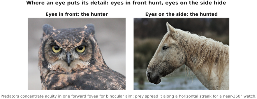
- Predators concentrate acuity in a single forward-pointing fovea and face their eyes forward, trading total field of view for the binocular overlap that judges distance to a strike. Cats, owls, and primates share this frontal, high-acuity plan. Raptors go furthest, with two foveae per eye: a deep central one aimed forward for maximum detail and a shallower one aimed sideways.
- Prey animals spread their acuity along a horizontal band, the visual streak, a ridge of high receptor density running side to side across the retina. Paired with eyes set on the sides of the head, it gives a grazer a sharp, panoramic view of the whole horizon, close to 360 degrees, without moving its eyes, so a predator anywhere on the skyline is caught. The streak lines up with the flat horizon of the open country these animals live in.
The old rule of thumb captures it: eyes in front, the animal hunts; eyes on the side, the animal hides (Figure 3.6.7). Either way the retina spends its fixed budget of receptors where ecology needs resolution, not evenly across the frame the way our cameras still do.
3.6.5 Seeing in the dark: sensitivity and its tricks⧉
The second axis, alongside where detail sits, is how little light an eye can work with. Nocturnal and deep-sea animals push sensitivity to places our own vision cannot follow, and every trick they use has a photographic twin.
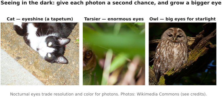
- Open the aperture. A larger pupil relative to focal length is a smaller f-number, and nocturnal eyes run remarkably fast: a cat's wide-open eye is around f/0.9, faster than almost any camera lens. Owls and the tarsier enlarge the whole eye to grow both the aperture and the image.
- Trade color and detail for photons. A rod-dominated retina, with many receptors pooled onto one nerve fiber (spatial summation), and a longer integration time (temporal summation, the eye's version of a slow shutter) both buy sensitivity by spending resolution. Nocturnal bees and the elephant hawkmoth summate so effectively that they see color by starlight, where our own color vision shut off long ago.
- Give each photon a second chance. The tapetum lucidum, a mirror behind the retina, reflects unabsorbed light back through the receptors, the source of the eyeshine in a flash photo. A cat's tapetum returns 10 to 20 percent of the light, a nocturnal gecko's 30 to 35 percent (Figure 3.6.8).
- Count single photons. At the extreme, a dark-adapted rod, in us as in a toad or a cockroach, can respond to a single photon, and the visual system fuses a handful of such events into a perception. The nocturnal dung beetle reads a cue 100 million times fainter than daylight, orienting by the faint band of the Milky Way and by the polarization of moonlight.
Some benchmarks of low-light vision, all far beyond a comfortable human threshold:
| Animal | Trick | Low-light feat |
|---|---|---|
| Cat | tapetum, ~f/0.9 pupil, rod-rich retina | works on roughly a sixth of the light we need |
| Tawny owl | huge tubular eyes, rod-rich retina | hunts by starlight under a closed canopy |
| Tarsier | eyes each larger than its brain | fully active on moonlight |
| Nocturnal helmet gecko | cone-based night vision, tapetum | tells colors apart by dim moonlight (~0.002 cd/m²) |
| Elephant hawkmoth | strong spatial/temporal summation | color vision in starlight |
| Dung beetle | polarization sensing, summation | steers by the Milky Way; reads moonlight polarization 10⁸× below daylight |
| Deep-sea fish | tubular eyes, stacked retinas | works on a few photons a kilometer down |
| Human (dark-adapted) | rod vision, ~f/2.1 pupil | detects a few photons; sees 6th-magnitude stars |
The lesson the noise chapter will later put in numbers is written across these animals: sensitivity is bought by spending something else, aperture, resolution, color, or speed, and evolution has tried every one of those trades.
3.6.6 Acuity: the finest detail, and how to sharpen it⧉
The counterpart to sensitivity is acuity: the finest detail an eye can resolve, set by how tightly its receptors sample the image and by the aperture and diffraction of its optics. The natural unit is the one an optical engineer uses, cycles per degree: how many black-and-white line pairs fit into one degree of the visual field before they blur to gray. A human fovea reaches roughly 60 cycles per degree, about one arcminute per line, the "20/20" line on the eye chart.
Two moves raise acuity, and they are the two a photographer already knows. Sample more finely: pack the receptors tighter, which runs up against their physical size and against diffraction. Make the image larger: a bigger eye is a longer focal length, and a longer focal length spreads the same scene across more receptors. Raptors combine both and add a third. An eagle carries cones several times denser than ours and a large eye for a long focal length, and its deep central fovea is shaped like a pit whose curved wall acts as a small diverging lens, magnifying the central image onto still more receptors, a built-in teleconverter. The result is the sharpest eye known, around 140 cycles per degree, roughly twice human acuity, which is how a wedge-tailed eagle reads a rabbit from a kilometer up.
At the other end, small eyes are hard against a wall. A compound eye's acuity is fixed by the angle between neighboring facets, and packing facets finer runs into diffraction almost at once; to reach human acuity a compound eye would have to be about a meter across, which is why an insect tops out near one cycle per degree. The jumping spider evades the limit with a narrow, telephoto principal eye whose long focal length and boosted central retina reach about 12 cycles per degree in a body a centimeter long (Figure 3.6.9).
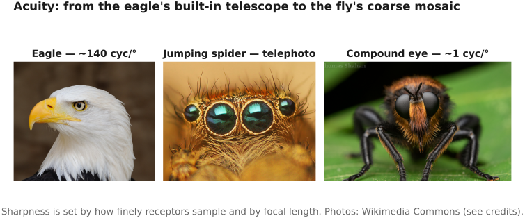
A camera, unlike an eye, samples uniformly, so one number describes it. The table below sets several animals against a modern phone's cameras and asks the reverse question a photographer cares about: what focal length would a 60-megapixel full-frame camera need, with a perfect lens, to sample as finely as each eye.
| Eye / camera | Visual acuity (cycles/°) | 60 MP full-frame focal length to match |
|---|---|---|
| Wedge-tailed eagle | ~140 | ~60 mm |
| Phone 5× telephoto (120 mm-eq, 12 MP)† | ~120 | (is a camera) |
| Human fovea | ~60 | ~22 mm |
| Phone main (24 mm-eq, 48 MP)† | ~55 | (is a camera) |
| Octopus | ~46 | ~14 mm |
| Ostrich | ~20 | any lens surpasses it |
| Phone ultrawide (13 mm-eq, 12 MP)† | ~19 | (is a camera) |
| Jumping spider | ~12 | any lens surpasses it |
| Cat | ~10 | any lens surpasses it |
| Goldfish | ~7 | any lens surpasses it |
| Rat | ~1 | any lens surpasses it |
| Honeybee | ~0.5 | any lens surpasses it |
| Housefly | ~0.1 | any lens surpasses it |
†Nominal pixel-sampling limit assuming a perfect lens; real phone optics and diffraction roughly halve it. The full-frame "focal length to match" puts a 60 MP sensor's pixels across a field narrow enough to sample at that acuity (a perfect, diffraction-free lens): a longer lens narrows the field and raises the sampled acuity. Below about 26 cycles per degree even a fisheye-wide full-frame lens over-samples the eye, so "any lens surpasses it."
Two things fall out. First, the phone in your pocket is not toy optics: its main camera already samples near the human fovea, and its 5× telephoto, on paper, out-resolves an eagle. Real lenses and diffraction pull that back, but the pixel grid is genuinely that fine. Second, the reach of a full-frame camera maps cleanly onto focal length: with a high-resolution sensor a normal 50 mm lens already out-samples the human eye, and it takes only a short 60 mm telephoto to match an eagle. To sample as coarsely as a horse or a cat you would need a lens wider than any that exists, which is a compact way of saying a modern camera out-resolves almost every eye in nature except the few sharpest birds.
3.6.7 Color vision: opsins remixed⧉
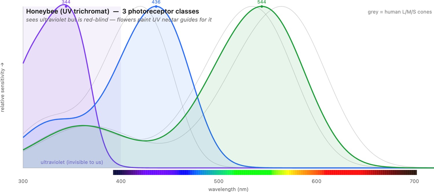
If the optics vary, color vision varies even more, because "color" is nothing but whichever set of opsins (the light-sensitive photopigment proteins) an animal happens to carry, and how it compares their outputs. Laid side by side, the photoreceptor spectral sensitivities of different animals make the diversity concrete (Figure 3.6.10): where we have three heavily-overlapping cones, a bee has three shifted into the ultraviolet, a bird or a fish has four, a dog has two, and a mantis shrimp has a dozen narrow ones spanning deep UV to red. Opsins are not even the whole story: many birds, reptiles, and fish also mount colored oil droplets in front of their photoreceptors, tiny filters that reshape a cone's sensitivity before the light ever reaches the pigment, a trick worth its own look at the end of the section. Three lessons stand out, all of them sharpening points made in Perceptual color and trichromatic vision.
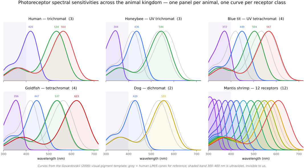
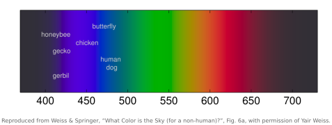
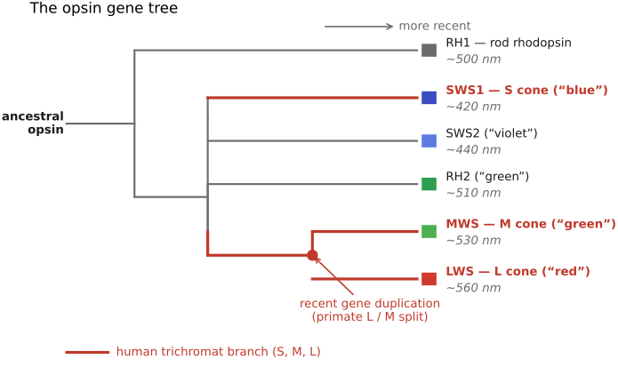
- More dimensions than three. Our trichromacy is not a ceiling. Many birds, reptiles, and fish are tetrachromats, with a fourth cone reaching into the ultraviolet (UV), they see patterns on flowers and plumage that are simply invisible to us. Most mammals, by contrast, are dichromats (red–green "color-blind" by our standard); primate trichromacy is re-evolved, the product of a recent gene duplication that split one pigment into our overlapping L and M cones, which is exactly why L and M overlap so heavily (see the opsin gene tree below). The lesson from the metamerism section of Perceptual color and trichromatic vision returns with teeth: every species is "color-blind" relative to some other, ourselves included, because every visual system projects an infinite spectrum onto a handful of numbers.
- More receptors do not mean more discrimination. The mantis shrimp carries roughly twelve to sixteen photoreceptor classes, and yet discriminates colors rather poorly in behavioural tests Thoen et al. 2014. The leading interpretation is that it does not compare channels the way a trichromat does (the comparison that the univariance discussion in Perceptual color and trichromatic vision showed is essential); instead it appears to recognize colors almost directly, reading off which receptor fires hardest. It is a working counterexample to "more channels = better color," and a reminder that color lives in the wiring, not just the pigments.
- Sampling axes we lack entirely. Some animals sense components of the plenoptic function we cannot. Pit vipers image thermal infrared with a separate pit organ, and UV-sensitive species push past our short-wavelength edge. Two further axes, the polarization of light and the speed at which the world is sampled in time, get their own sections below. Each species samples a different slice of the same physical world.
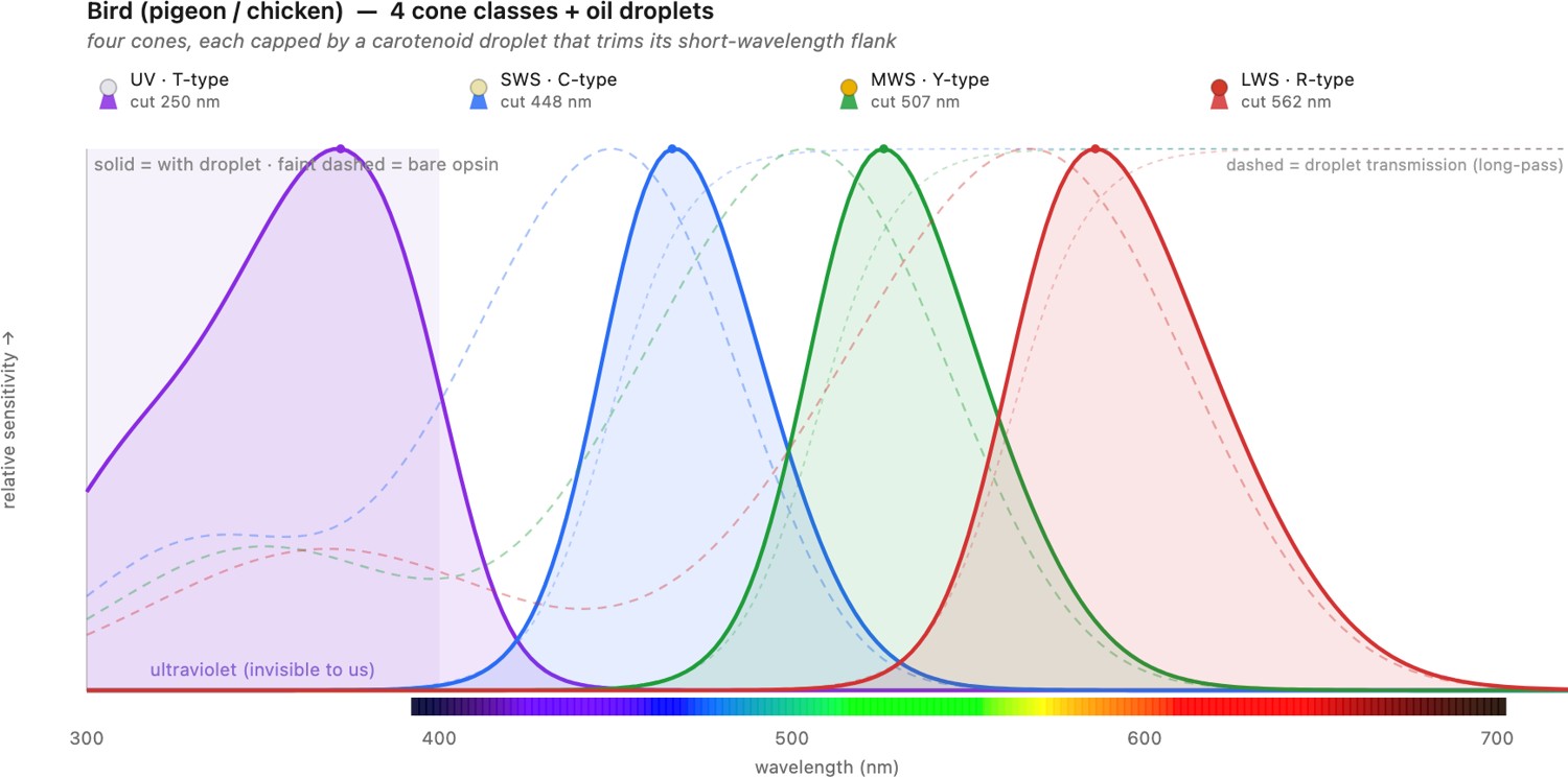
Those oil droplets repay the closer look promised earlier, because they are the clearest case of an eye tuning its color sense without touching its pigments. A bird's retina is studded with them in a fixed palette: a deep red droplet in front of each long-wavelength cone, a yellow-orange one ahead of the mid cones, and paler, nearly clear ones over the short-wavelength and ultraviolet cones. Each is a carotenoid long-pass filter that shaves the short-wavelength flank off the cone beneath it, so the four channels come out far narrower and less overlapped than the raw opsins would leave them, part of why birds out-discriminate us across much of the spectrum while keeping a clean ultraviolet channel. Turtles carry an even more vivid set, some lizards and the dipnoan lungfishes have them too, and the pigeon and chicken retinas are the textbook maps. It amounts to a color-filter array built one droplet per receptor, the biological ancestor of the dye mosaic on a camera sensor: tune the spectral response by subtracting light on the way in, rather than by evolving a new photopigment (Figure 3.6.14).
All of this divergence is written in the opsin gene tree. From a single ancestral opsin, gene duplication and loss produced the pigment classes that different animals draw on, and our own re-evolved L/M split is one recent twig on it, which is why those two cones overlap so heavily (Figure 3.6.12). An interactive explorer lets you dial up any species' photoreceptor curves against our own and watch its world of color reach past ours (Figure 3.6.13). The phylogeny is developed in the "Evolution of Eyes" chapter of Vision (Cambridge) Vision, "Evolution of Eyes".
3.6.8 Polarization: the axis we are blind to⧉
Light carries more at each direction than an intensity and a spectrum. Its electric field oscillates in a particular orientation, and that orientation, the polarization, is a dimension our own eyes throw away almost entirely (recall polarization from Light and optics). Sunlight leaves the sun unpolarized, but the world polarizes it: Rayleigh scattering in the sky, reflection off water and leaves and cuticle, and scattering underwater all stamp a partial polarization onto the light that a suitable eye can read. Many animals read it, and put it to uses we can only approach with a filter screwed onto a lens.
A compass in the sky. Rayleigh scattering lays a fixed pattern of polarization across the blue sky, bands that wheel around the sun and move with it through the day. Insects read the pattern as a compass. A foraging honeybee or a desert ant holds its heading by the sky's polarization even when the sun itself is hidden behind a cloud or a dune, using a specialized strip of the eye, the dorsal rim area, whose receptors are tuned to the angle of polarization. It is celestial navigation with no stars and no clock, run off a patch of blue.
Finding water, and cutting glare. Light reflected from a horizontal surface comes back strongly polarized in the horizontal direction, the very fact a photographer uses when a polarizing filter kills the glare on a pond or a windshield. Water beetles, dragonflies, and mayflies find water by exactly that horizontal polarization, diving toward any surface that reflects it, which is why they are now regularly fooled into laying eggs on car roofs, asphalt, and solar panels. The animal carries a permanent polarizer and a brain wired to what it reveals.
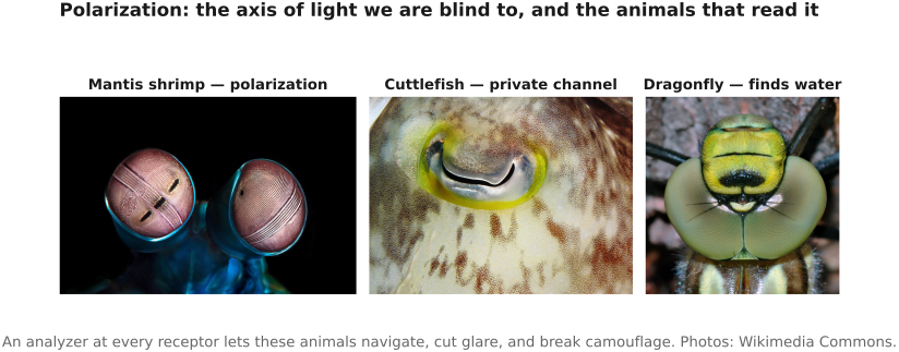
A private, high-contrast channel underwater. Below the surface, where color washes out with depth but polarization persists, the cephalopods excel. Octopus, cuttlefish, and squid are colorblind, yet their polarization vision is superb, and they lean on it where color fails: to lift a silvery, transparent, or countershaded animal out of a flat background, since the camouflage that beats a brightness-and-color eye still disturbs the polarization of the light passing through or bouncing off it. Cuttlefish may even signal to one another with polarized patterns on their skin, a channel a colorblind predator cannot read. The mantis shrimp goes furthest, the only animal known to sense circular polarization as well as linear, perhaps for just such a private band (Figure 3.6.15).
The reason this is largely an invertebrate gift is in the receptor. An invertebrate photoreceptor is built from tightly packed microvilli, tubes whose aligned pigment molecules absorb light polarized along their axis and pass the rest, so a single receptor is already a polarization analyzer. Our cones use a different, ciliary construction whose pigment tumbles freely and averages polarization away, which is why we are nearly blind to it, save for the faint "Haidinger's brush" a few people learn to glimpse in a clear blue sky.
For imaging, polarization is the axis the camera left on the table for a century. A conventional sensor is as polarization-blind as we are, and a screw-on polarizer samples only one angle at a time. The animals' trick, an analyzer at every receptor, is exactly what a modern polarization sensor now copies, tiling the pixel grid with micro-polarizers at four orientations so one exposure records the polarization everywhere. That reopens the same uses evolution found: cut glare and see into water, tell materials apart by how they polarize a reflection, and recover surface shape from the polarization of the light a scene returns (shape from polarization). The eye that steers by the sky and the camera that maps stress in a windshield are reading the same neglected axis of the light.
3.6.9 Temporal vision: flicker, and motion first⧉
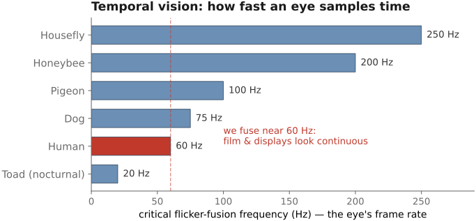
An eye samples the world in time as well as in space, and animals differ as sharply in their frame rate as in their acuity. The summarizing number is the critical flicker fusion frequency: the rate above which a flickering light fuses into a steady glow. Ours is around 50 to 60 hertz, which is why film at 24 frames (each flashed twice) and a display refreshing at 60 hertz or more read as continuous motion, and why we do not see the mains-driven flicker of the lights around us.
That number tracks how an animal lives. A fast flier sees time in slow motion: a housefly does not fuse flicker until well above 250 hertz, so our quickest swat arrives, to the fly, as a slow and readable approach, and a television would look to it like a slideshow. Birds sit near 100 hertz and above; a film made to look smooth to a bird would need far more frames than one made for us. The cost is the shutter speed. A high fusion frequency means a short integration time, which gathers few photons, so nocturnal and deep-sea animals run slow, pooling light over long exposures at the price of smearing anything that moves, the same sensitivity-versus-motion-blur trade a photographer weighs at every shutter setting. Body size and metabolism set the scale: small, fast-burning animals carry the highest fusion frequencies and, in a real sense, experience time as flowing more slowly than we do (Figure 3.6.16).
The other half of temporal vision is that detecting motion is older, and often more urgent, than resolving form. A moving predator or a moving meal matters more than a static scene, and many eyes compute motion first, sometimes almost to the exclusion of everything else. The classic demonstration is the frog. Lettvin and colleagues, recording from the frog's retina, found cells that fire only for small, moving, convex edges, the "bug detectors," while the stationary world barely registers; a frog ringed by freshly killed, motionless flies will starve. A toad strikes only at moving, worm-shaped stimuli, and freezes if they stop. The machinery behind this is a motion detector wired directly into the retina: direction-selective cells that respond to one direction of drift and not its reverse, described since the 1950s by the Hassenstein-Reichardt correlator, which compares a receptor's signal against a delayed copy of its neighbor's. It is the same computation the book will later build as optical flow. Even in us, motion rules the periphery: we catch a flicker of movement at the edge of sight and swing the fovea to it, form arriving only after motion has raised the alarm.
The engineering echo is the sharpest in the chapter. The event camera, or dynamic vision sensor, is motion-first vision cast in silicon: each pixel reports the instant its own brightness changes, asynchronously, with microsecond latency, rather than the whole array marching out frames on a clock. It captures fast motion with no blur, emits almost no data when nothing moves, and reacts in microseconds, exactly the fly-eye bargain, and its output is a stream of moving edges a frog would recognize. Motion-first vision, ancient in the retina, is being rebuilt in the sensor.
The throughline of the whole chapter, now stated across species: color and vision are not read off the world, they are computed from it, projected through whatever opsins an eye carries, re-coded by whatever wiring sits behind them, adapted, and inferred. Human vision is one particular such computation, and, ranked across the axes this chapter has toured, a capable generalist rather than a record-holder: our daylight acuity sits near the animal ceiling (though a hawk still out-resolves us), our color is solid trichromacy (a bird or a mantis shrimp carries far more channels), our low-light sensitivity is genuine but modest (a cat works on a sixth of the light we need), our flicker rate is middling next to a fly's, and polarization and the ultraviolet we give up altogether. No single axis is won outright, yet none is badly lost, which is a fair description of a generalist eye. Every later chapter on color, encoding, and image quality is an attempt to live within, or exploit, the constraints our version of that biological processor imposes.